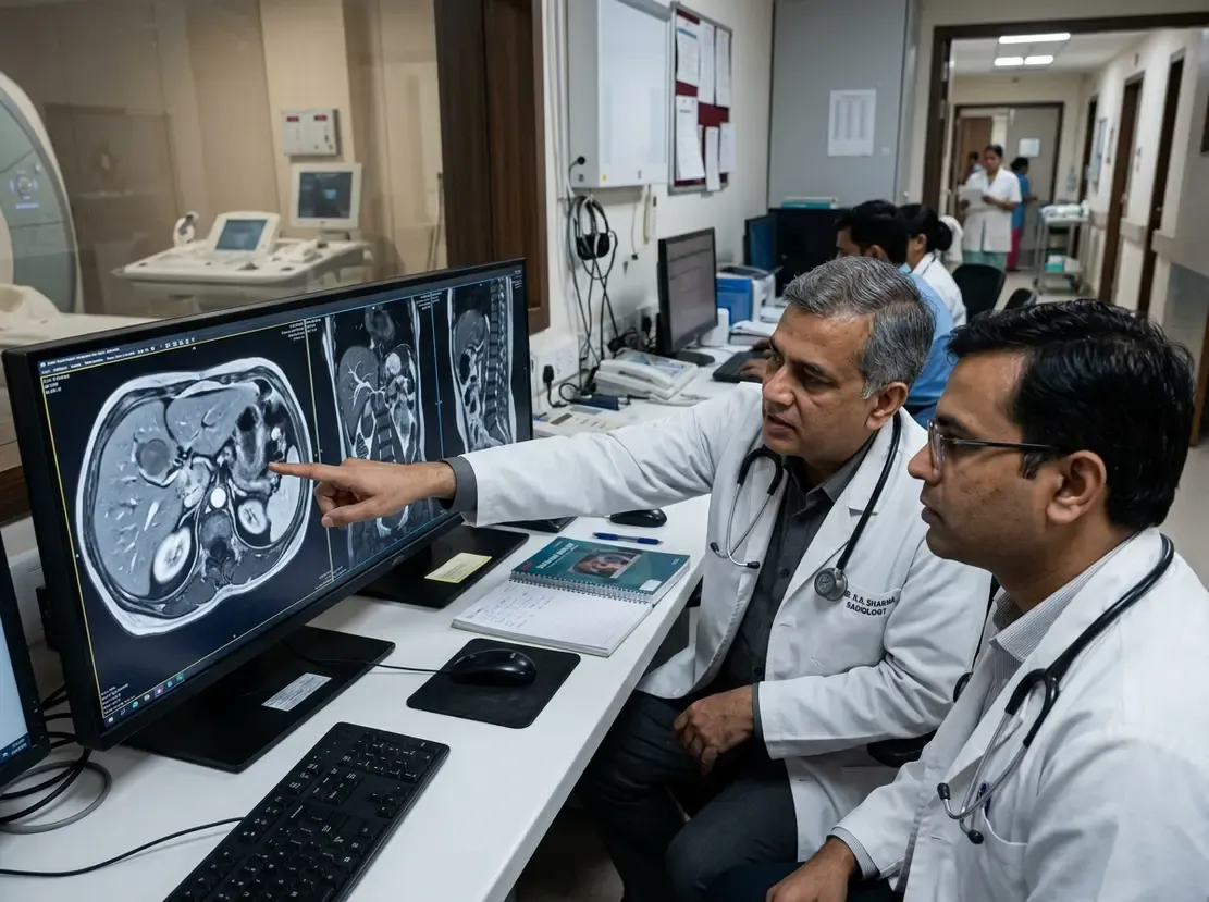
Is Ellipsoid Prostate Volume Reliable for PSA Density?

Accurate prostate volume assessment plays a pivotal role in evaluating prostate cancer risk. Clinicians frequently use multiparametric MRI to measure gland dimensions and calculate prostate-specific antigen density (PSAd). However, manual segmentation of whole-gland MRI slices requires substantial time and expertise. Consequently, most urologists and radiologists rely on the faster ellipsoid approximation formula.
Manual Segmentation vs Ellipsoid Prostate Volume Assessment
A prospective study examined whether the standard ellipsoid formula matches manual planimetric contouring. Specifically, investigators analyzed 328 patients who underwent 3-Tesla multiparametric MRI for suspected prostate cancer. Radiologists calculated reference volumes by manually tracing all axial T2-weighted slices. Meanwhile, they derived ellipsoid volumes using the standard PI-RADS equation, multiplying three orthogonal diameters by 0.52.
Overall, the ellipsoid approximation demonstrated excellent statistical agreement with manual segmentation. The intraclass correlation coefficient reached 0.985, showing a modest mean absolute difference of 5.5 cc. Therefore, standard three-dimensional measurements reliably reflect actual gland dimensions in routine clinical environments.
Impact on Large Glands and Individualized Weighting
Although the ellipsoid formula performed well overall, discrepancies widened in larger prostate glands. In prostates exceeding 80 cc, the geometric ellipsoid formula tended to underestimate gland volume. To address this variance, researchers tested whether individualized weighting factors improved calculation accuracy.
Statistical models analyzed variable weighting coefficients ranging from 0.50 to 0.54. Nonetheless, fine-tuning these coefficients provided no meaningful improvement over the standard 0.52 factor. Thus, modifying the standard mathematical constant fails to offer tangible diagnostic advantages in practice.
Implications for PSA Density and Clinical Decisions
Importantly, volume discrepancies did not impair the diagnostic performance of PSA density. Across established clinical thresholds of 0.10, 0.15, and 0.20 ng/mL/cc, the ellipsoid method maintained high diagnostic fidelity. It achieved sensitivities between 0.86 and 0.95 alongside specificities of 0.88 to 0.97 compared with manual segmentation.
Consequently, clinicians can confidently use the rapid PI-RADS ellipsoid method during patient triage. Urologists frequently use PSA density to guide biopsy choices and monitor men on active surveillance. Because manual contouring remains labor-intensive, the ellipsoid shortcut provides dependable risk stratification without delaying clinical workflows.
Frequently Asked Questions
Q1: Why does the ellipsoid formula underestimate large prostate volumes?
As benign prostatic hyperplasia progresses, the transitional zone enlarges irregularly. This nodular expansion distorts the spherical shape, making large glands deviate from ideal ellipsoid geometry.
Q2: Does volume underestimation alter PSA density risk categorization?
Despite minor volume underestimates in glands over 80 cc, PSA density thresholds remain robust. The ellipsoid calculation reliably preserves high sensitivity and specificity for clinical decision-making.
Q3: Should radiologists adjust the 0.52 weighting factor for different gland sizes?
No. Research demonstrates that adjusting the weighting factor between 0.50 and 0.54 yields no significant clinical benefit over the standard 0.52 coefficient.
References
- Gluesing L et al. MRI-derived prostate volume assessment: manual segmentation versus two-axis ellipsoid approximation and its implications for PSA density. Eur Radiol. 2026 Sep 28. doi: 10.1007/s00330-026-12900-1. PMID: 42803939.
- Ponsiglione A, Stanzione A, Vitale R, et al. Prostate Volume Estimation on MRI: Accuracy and Effects of Ellipsoid and Bullet-Shaped Measurements on PSA Density. Acad Radiol. 2021;28(6):805-812.
- Nandalur KR, Baccouche H, Hryhorczuk A, et al. Which measurement method should be used for prostate volume for PI-RADS? A comparison of ellipsoid and segmentation methods. Clin Imaging. 2022;81:41-45.