CT vs MRI: Choosing the Best Pancreatic Cancer Imaging

Clinical Importance of Pancreatic Cancer Imaging
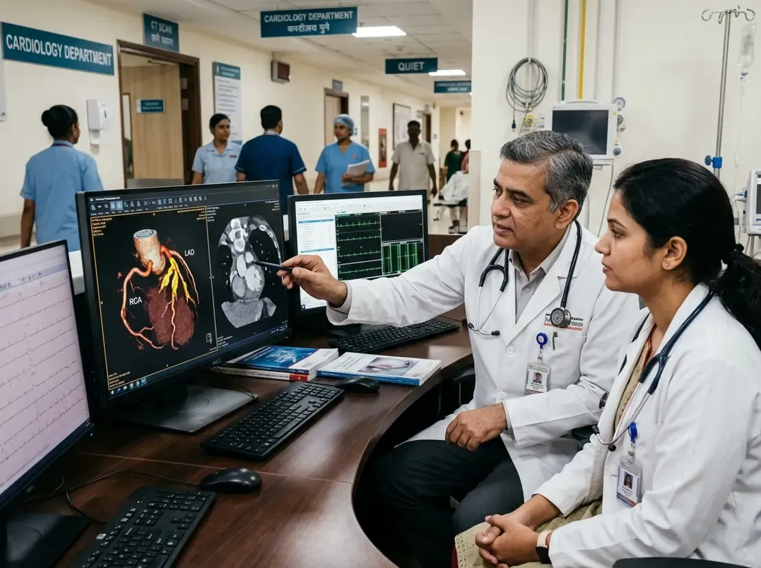
Pancreatic ductal adenocarcinoma remains an aggressive malignancy with a narrow therapeutic window. Consequently, accurate pancreatic cancer imaging is vital for identifying candidates for curative surgical resection. Clinicians face persistent challenges when choosing among computed tomography, magnetic resonance imaging, and endoscopic ultrasound. Therefore, a recent meta-analysis compared these modalities to establish clear diagnostic benchmarks.
Primary Tumor Detection and Local Staging
The systematic review analyzed forty direct comparative studies. Interestingly, computed tomography, magnetic resonance imaging, and endoscopic ultrasound showed comparable diagnostic performance for detecting primary tumors. None of the three modalities demonstrated clear superiority in identifying pancreatic ductal adenocarcinoma lesions.
However, each modality excels in distinct clinical aspects. For instance, endoscopic ultrasound demonstrated higher sensitivity for detecting regional lymph node involvement than computed tomography or positron emission tomography. Thus, clinicians can leverage endoscopic ultrasound when local nodal status dictates neoadjuvant therapy decisions, which is particularly valuable for specialists pursuing advanced gastrointestinal imaging expertise.
Vascular Invasion and Resectability Assessment
Surgical planning depends directly on vascular involvement and overall resectability. In this domain, computed tomography outperformed magnetic resonance imaging when evaluating major vascular invasion. Furthermore, computed tomography achieved a higher specificity of 0.88 for assessing resectability, compared to 0.63 for magnetic resonance imaging.
Accordingly, surgeons rely on computed tomography to determine whether a tumor encases major peripancreatic vessels. This high specificity prevents clinicians from declaring resectable tumors as unresectable. Consequently, dedicated pancreatic-protocol computed tomography remains the cornerstone for surgical staging.
Detecting Liver Metastases: MRI Outperforms CT
Liver metastases immediately change disease management from curative resection to systemic chemotherapy. In the meta-analysis, magnetic resonance imaging showed superior pooled sensitivity of 0.89 and specificity of 0.91 for identifying liver metastases. In contrast, computed tomography demonstrated a pooled sensitivity of only 0.60 and specificity of 0.84.
Therefore, contrast-enhanced abdominal computed tomography often misses small occult liver metastases. Adding a liver magnetic resonance imaging protocol before surgery can rule out subtle distant lesions. As a result, this targeted approach saves patients from unnecessary laparotomies.
Clinical Recommendations for Indian Oncologists
In Indian clinical settings, resource allocation and cost represent critical considerations. Clinicians should initially obtain high-resolution multiphase computed tomography to stage disease and rule out major vascular encasement. Moreover, computed tomography effectively surveys chest and abdominal disease burden.
However, oncologists should strongly consider liver magnetic resonance imaging before undertaking major pancreatic surgery. This step provides crucial assurance against microscopic or sub-centimeter hepatic deposits. Additionally, endoscopic ultrasound remains an invaluable problem-solving tool for doubtful lymph nodes or tissue confirmation, aligning with competencies expected in a comprehensive oncology training program.
Frequently Asked Questions
Q1: Which imaging modality best assesses pancreatic cancer resectability?
Multiphase computed tomography offers the highest specificity for assessing surgical resectability and vascular invasion. Consequently, it serves as the preferred first-line imaging modality for resectability evaluation.
Q2: Why should clinicians order a liver MRI before pancreatic resection?
Magnetic resonance imaging achieves significantly higher sensitivity and specificity for detecting liver metastases than computed tomography. Therefore, it reliably uncovers small liver metastases that could preclude curative resection.
Q3: What role does endoscopic ultrasound play in pancreatic staging?
Endoscopic ultrasound provides excellent sensitivity for detecting regional lymph node metastasis. Furthermore, it enables fine-needle biopsy for definitive histological diagnosis.
References
- Rezuș II et al. Imaging-based diagnosis of pancreatic cancer: a systematic review and meta-analysis of direct comparative studies. Eur Radiol. 2026 Oct 01. doi: 10.1007/s00330-026-12890-0. PMID: 42821086.
- Kalinov T. Comparative analysis of imaging modalities for pancreatic cancer detection: A comprehensive review of diagnostic performance. Scr Sci Med. 2025;57(2):15-24.
- Conroy T, Pfeiffer P, Vilgrain V, et al. ESMO Clinical Practice Guideline: Pancreatic Cancer. Ann Oncol. 2023;34(9):787-802.