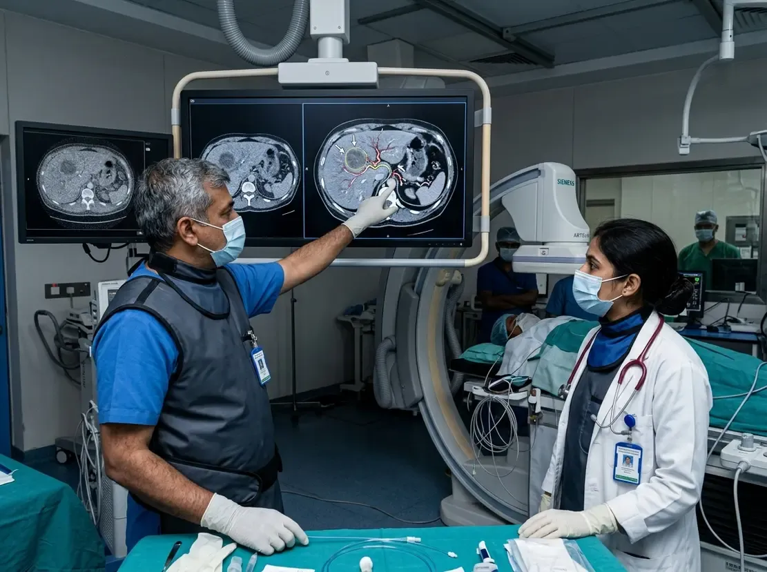
DCE-MRI Predicts Early Pancreatic Cancer Therapy Success

Pancreatic ductal adenocarcinoma remains one of the most lethal malignancies worldwide. However, clinicians often face major challenges when assessing early treatment efficacy using standard CT scans. A new prospective study demonstrates that quantitative DCE-MRI can accurately predict chemotherapy response after only seven weeks of therapy. Consequently, this innovative functional imaging method offers a significant leap forward over conventional anatomical imaging modalities.
Role of Quantitative DCE-MRI in Pancreatic Cancer
Conventional CT relies primarily on morphologic measurements to gauge therapeutic efficacy. Unfortunately, structural tumor regression often takes several months to appear in dense desmoplastic tumors. In contrast, quantitative DCE-MRI evaluates microvascular perfusion and vessel permeability much earlier in the treatment course. In this multicenter study, researchers prospectively evaluated 42 patients before chemotherapy and seven weeks after starting systemic treatment. Furthermore, investigators derived 11 pharmacokinetic parameters to track microvascular changes over time.
Key Study Findings and Error Correction
The investigators discovered that early changes in vascular transfer constant values significantly distinguished responders from non-responders. Initially, uncorrected imaging protocols achieved approximately 80% predictive accuracy. However, scanner-dependent measurement variations can introduce noticeable errors into pharmacokinetic calculations. To resolve this challenge, researchers utilized a point-of-care portable perfusion phantom concurrently during scans. After phantom-based calibration, the predictive accuracy surged to 98% with the extended Tofts model. In sharp contrast, standard RECIST CT assessments at eight weeks detected response in merely 17% of patients.
Clinical Implications for Oncology Practice
These findings carry substantial clinical relevance for daily oncological decision-making. First, early identification of non-responders allows clinicians to modify ineffective chemotherapy regimens promptly. Second, timely clinical intervention spares patients from unnecessary drug toxicities. Therefore, phantom-corrected imaging facilitates truly personalized therapeutic strategies for pancreatic cancer. As a result, patients can receive optimized care pathways much earlier.
Frequently Asked Questions
Q1: Why is quantitative DCE-MRI superior to standard CT for early PDAC evaluation?
Standard CT measures gross anatomical shrinkage, which often develops slowly in pancreatic tumors. In contrast, quantitative DCE-MRI measures functional vascular permeability changes that emerge within weeks of starting systemic therapy.
Q2: How does the portable perfusion phantom improve MRI accuracy?
The portable perfusion phantom corrects scanner-dependent measurement drift. Consequently, it standardizes pharmacokinetic parameters across imaging sessions and raises predictive accuracy up to 98%.
References
- Holland MD et al. Quantitative dynamic contrast-enhanced magnetic resonance imaging for early assessment of treatment response in pancreatic ductal adenocarcinoma. Eur Radiol. 2026 Aug 22. doi: 10.1007/s00330-026-12810-2. PMID: 42631739.
- Kim H, Morgan DE, Schexnailder P, et al. Accurate therapeutic response assessment of pancreatic ductal adenocarcinoma using quantitative dynamic contrast-enhanced magnetic resonance imaging with a point-of-care perfusion phantom: a pilot study. Invest Radiol. 2019;54(1):16-22.
- Tang W, Liu W, Li HM, et al. Quantitative dynamic contrast-enhanced MR imaging for the preliminary prediction of the response to gemcitabine-based chemotherapy in advanced pancreatic ductal carcinoma. Eur J Radiol. 2019;121:108734.