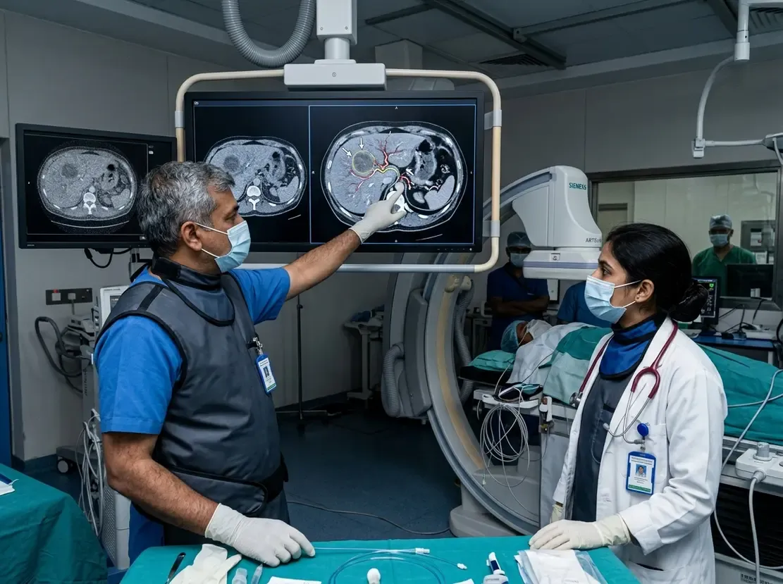
Spectral CT Accurately Rules Out Pancreatic Malignancies

The Clinical Dilemma of Intrapancreatic Accessory Spleen
Differentiating an intrapancreatic accessory spleen from hypervascular pancreatic neoplasms poses a major diagnostic dilemma for clinicians. In clinical practice, benign ectopic splenic tissue often mimics neuroendocrine tumors on conventional imaging scans. Consequently, patients frequently face invasive workups or unnecessary pancreatic resections. Fortunately, novel dual-energy computed tomography methods now offer distinct quantitative insights. Therefore, radiologists can accurately identify tissue composition without subjecting patients to multiple scan phases.
How Spectral CT Evaluates Pancreatic Tissue
A recent landmark study evaluated single-phase portal-venous spectral CT in eighty-five patients with hypervascular pancreatic lesions. Specifically, investigators evaluated thirty-four cases of intrapancreatic accessory spleen and fifty-one neoplasms. The team analyzed quantitative parameters across multiple spectral reconstructions. These reconstructions included virtual monochromatic images, virtual non-contrast images, iodine concentration maps, and effective atomic number maps. In addition, researchers calculated the relative lesion-to-spleen ratio for each parameter. Because splenic tissue maintains uniform physical characteristics, values closer to zero indicated true splenic origin.
Quantitative Findings and Diagnostic Accuracy
The quantitative analysis revealed significant diagnostic differences between benign lesions and tumors. First, relative lesion-to-spleen ratios were consistently lower in splenic tissue across all spectral maps. Furthermore, virtual monochromatic images at low photon energies achieved remarkable discriminative power. At forty kiloelectronvolts, the technique yielded an area under the curve exceeding 0.90. Moreover, effective atomic number and iodine concentration maps provided exceptional diagnostic fidelity. When three independent radiologists evaluated pancreatic tail lesions, adding spectral maps improved their pooled diagnostic accuracy significantly. Consequently, inter-observer agreement increased markedly among reading radiologists.
Clinical Implications for Practice
These findings offer substantial practical advantages for oncologic and surgical decision-making. Single-phase portal-venous imaging reduces cumulative radiation exposure compared to multiphasic CT protocols. However, the spectral data still provides robust material decomposition and tissue characterization. Thus, clinicians can confidently exclude malignancy and prevent unnecessary surgical distal pancreatectomy. Ultimately, incorporating spectral CT into routine abdominal imaging safeguards patients from substantial surgical morbidity.
Frequently Asked Questions
Q1: Why is an intrapancreatic accessory spleen frequently mistaken for a neoplasm?
An accessory spleen shares similar arterial hypervascularity and contrast enhancement patterns with small neuroendocrine tumors on conventional CT.
Q2: How does spectral CT differentiate splenic tissue from pancreatic tumors?
Spectral CT analyzes iodine maps, monochromatic energy levels, and effective atomic numbers to demonstrate tissue equivalence with the native spleen.
Q3: What clinical advantage does single-phase portal-venous spectral imaging provide?
It provides high diagnostic certainty while eliminating multiphasic contrast runs, which lowers the patient's overall radiation exposure.
References
- Nagayama Y et al. Spectral imaging for differentiating intrapancreatic accessory spleen from hypervascular pancreatic neoplasms using single-phase post-contrast CT. Eur Radiol. 2026 Oct 02. doi: 10.1007/s00330-026-12924-7. PMID: 42825957.
- Jang KM, Kim SH, Lee SJ, et al. Differentiation of an intrapancreatic accessory spleen from a small (<3-cm) solid pancreatic tumor: value of diffusion-weighted MR imaging. Radiology. 2013;266(1):159-167.
- Kim J, Lee JM, Han JK, et al. Diffusion-weighted MRI: usefulness for differentiating intrapancreatic accessory spleen and small hypervascular neuroendocrine tumor of the pancreas. Acta Radiol. 2014;55(10):1157-1165.