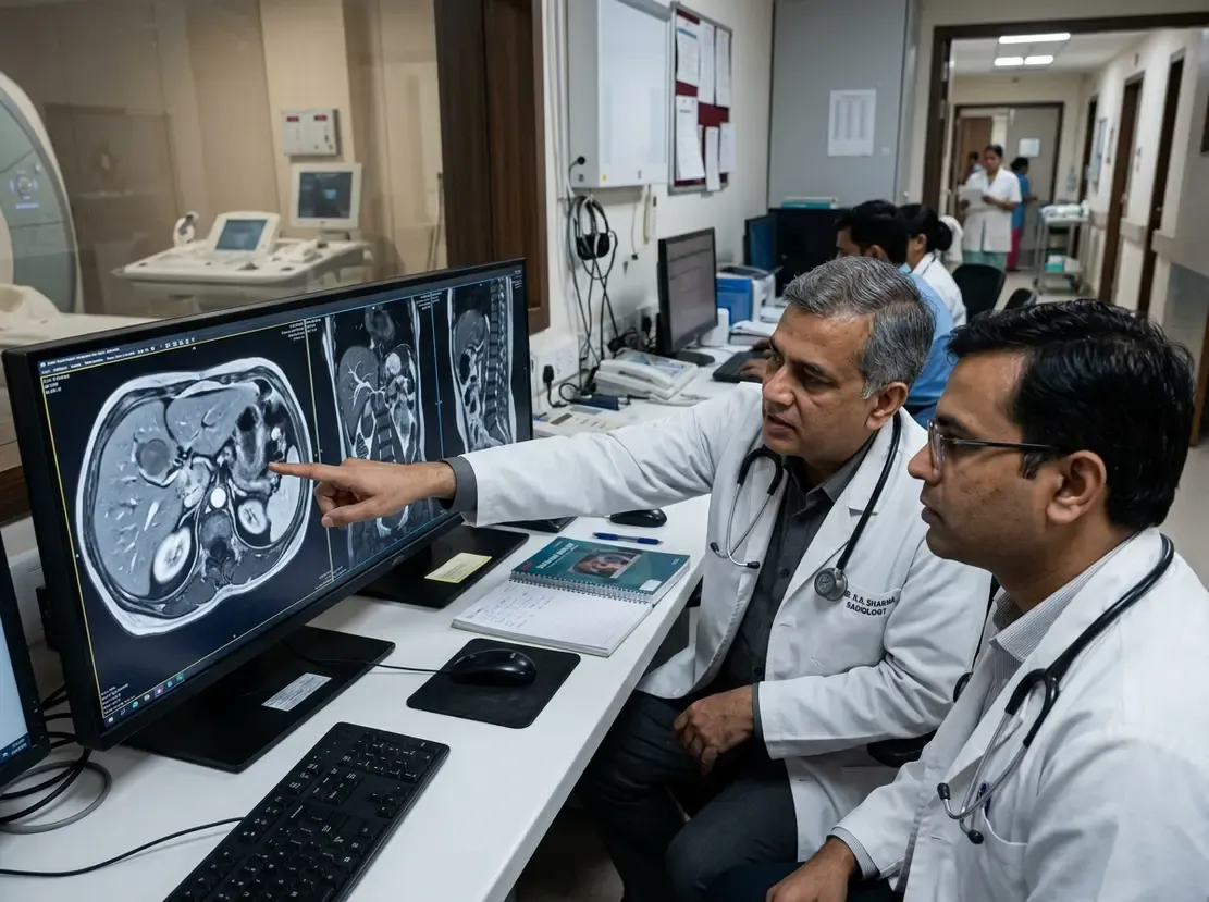
Standardizing Sacroiliac Joint CT Reporting in SpA
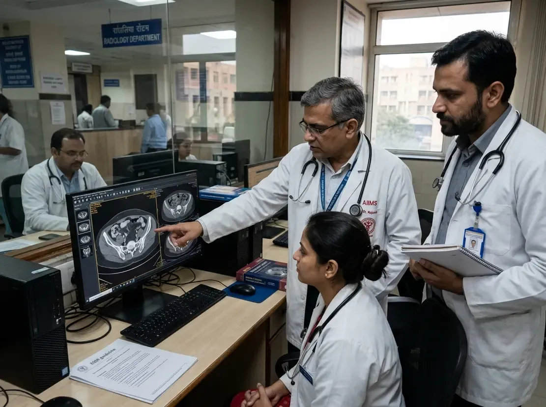
Accurate evaluation of spondyloarthritis demands objective and reproducible imaging standards. Consequently, the European Society of Musculoskeletal Radiology (ESSR) recently addressed significant variations in sacroiliac joint CT reporting. This multicenter initiative establishes clear morphological criteria for identifying structural changes. As a result, clinicians and radiologists gain a shared diagnostic vocabulary.
The Need for Sacroiliac Joint CT Reporting Standards
Computed tomography offers exceptional spatial resolution for cortical bone and joint architecture. However, radiologists frequently encounter ambiguous definitions when reporting axial spondyloarthritis. Variability in terminology often creates diagnostic confusion between degenerative changes and inflammatory lesions. Therefore, the ESSR Arthritis Subcommittee initiated a modified Delphi study.
A core panel of twelve international experts initially drafted proposed lesion definitions. Subsequently, a wider panel of up to 48 musculoskeletal radiologists evaluated these criteria over three successive rounds. The panel required a 75% agreement threshold and a score of at least 8 on a 10-point scale. Ultimately, consensus rates ranged between 76% and 96% across all selected definitions. These rigorous thresholds confirm strong expert alignment across international institutions.
Seven Consensus Definitions for Structural Lesions
The Delphi consensus finalized standardized definitions for seven structural sacroiliac joint abnormalities. First, experts defined erosions as distinct cortical bone defects with loss of subchondral bone plate. Second, the panel characterized pseudo-widening as apparent joint space widening resulting from extensive subchondral erosion. Third, subchondral sclerosis reflects increased bone density extending beyond normal physiological limits.
Additionally, the panel included SI joint space narrowing, defined as localized or generalized reduction in joint width. Furthermore, definitions for bone bud and ankylosis reached decisive approval. A bone bud represents early osseous bridging, whereas ankylosis indicates complete fusion across the joint space. Interestingly, the committee retained backfill as a recognizable structural lesion on CT. In contrast, the experts voted to exclude fat metaplasia because CT demonstrates limited sensitivity for marrow fat.
Clinical Impact and the Road to SIJ-RADS
Standardizing structural lesion terminology creates immediate advantages for everyday clinical practice. First, clear definitions reduce interobserver variability among radiologists. Consequently, clinicians receive far more reliable imaging assessments during diagnostic workups. Moreover, these harmonized definitions facilitate early discrimination between inflammatory sacroiliitis and mechanical stress.
Most importantly, these seven definitions provide the foundation for a future CT-based SIJ-RADS classification system. Similar to BI-RADS and LI-RADS, a standardized reporting and data system will streamline clinical communication. Thus, treating physicians can stratify sacroiliitis severity with greater confidence and initiate timely targeted therapy.
Frequently Asked Questions
Q1: Why did the ESSR panel exclude fat metaplasia from the CT lesion definitions?
The panel excluded fat metaplasia because computed tomography has limited sensitivity for subtle bone marrow fat changes compared to MRI.
Q2: What is the clinical significance of distinguishing bone buds from ankylosis?
Bone buds represent initial focal bony bridges across the joint, whereas ankylosis signifies established, complete osseous bridging across the articular space.
Q3: How do standardized CT lesion definitions assist in creating SIJ-RADS?
Uniform definitions establish objective grading criteria, facilitating standardized risk categories in a future SIJ-RADS scoring system.
References
- Korteweg MA et al. Standardized reporting of sacroiliac joints in spondyloarthritis on CT: modified Delphi consensus on lesion definitions from the ESSR-Arthritis Subcommittee. Eur Radiol. 2026 Sep 29. doi: 10.1007/s00330-026-12889-7. PMID: 42809116.
- Rennie WJ, Cotten A, Jurik AG, et al. Standardized reporting of spine and sacroiliac joints in axial spondyloarthritis MRI: from the ESSR-Arthritis Subcommittee. Eur Radiol. 2025;35(1):15-28. doi: 10.1007/s00330-024-10892-z.
- Diekhoff T, Hermann KGA, Greese J, et al. Low-dose computed tomography of the sacroiliac joints in axial spondyloarthritis: an alternative to MRI? Ann Rheum Dis. 2022;81(4):534-540.