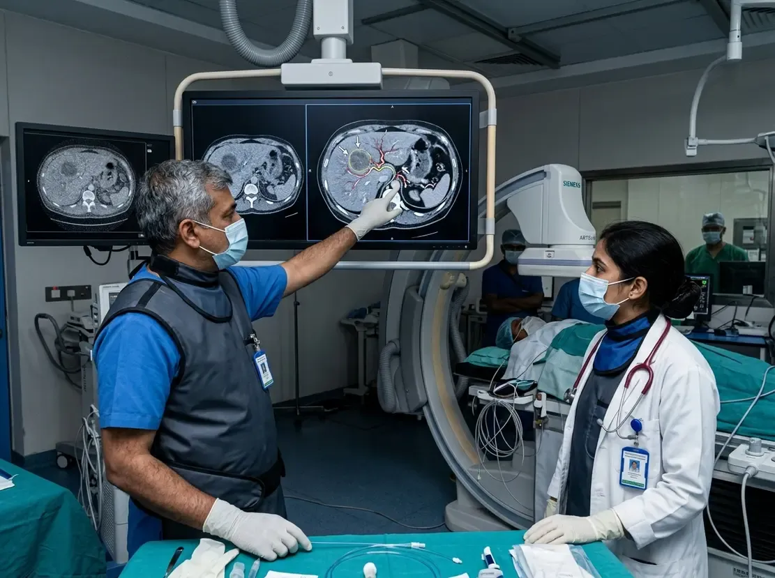
Spectral CT Predicts Gastric Cancer Therapy Response

Evaluating Gastric Cancer with Spectral CT Habitat Analysis
Oncologists constantly seek accurate tools to evaluate treatment response in gastric cancer patients. Therefore, non-invasive imaging techniques have gained significant interest in modern oncology. Specifically, spectral CT habitat analysis represents a cutting-edge approach to examining tumor heterogeneity. This technique extracts multi-parametric data from iodine maps to identify diverse perfusion patterns within a tumor. Indeed, assessing these subregions helps clinicians predict how well a patient will respond to neoadjuvant therapy. Consequently, physicians can tailor treatment plans and improve patient survival rates significantly.
Mapping Perfusion Habitats and Heterogeneity
Recently, researchers conducted a retrospective two-center study to evaluate this predictive model. First, the team analyzed pre-treatment spectral CT scans from 151 patients with gastric cancer. Additionally, they categorized the patients into responders and non-responders based on their tumor regression grade. Next, they clustered tumor voxels on venous-phase iodine maps into three distinct perfusion habitats. These three habitats represented areas of high, middle, and low tumor perfusion. Importantly, responders demonstrated a higher volume fraction of low-perfusion habitats compared to non-responders. Furthermore, responders exhibited a lower intratumoral heterogeneity score, which indicates a more uniform tumor microenvironment. Ultimately, the low-perfusion habitat volume fraction and the heterogeneity score proved to be independent predictors of response.
Clinical Benefits for Gastric Cancer Care
Gastric cancer poses a significant health challenge across many regions of India. Because early prediction of therapy response remains difficult, this novel imaging method offers a major clinical advantage. Specifically, identifying non-responders before surgery allows oncologists to modify ineffective regimens early. This proactive strategy saves valuable time and prevents unnecessary drug toxicities for the patient. Moreover, the dual-phase spectral CT scans utilize standard contrast protocols that many Indian diagnostic centers already perform. As a result, clinical teams can integrate this software-based habitat analysis without purchasing expensive new equipment. Finally, this non-invasive tool can assist surgical planning and optimize the timing of gastrectomy.
Frequently Asked Questions
Q1: What is spectral CT habitat analysis?
This method clusters tumor subregions into distinct habitats using iodine maps. Consequently, it reveals spatial variations in tumor blood flow.
Q2: How does this method predict treatment response?
Generally, responders show a higher volume fraction of low-perfusion habitats. Additionally, they exhibit lower heterogeneity scores before neoadjuvant therapy starts.
Q3: Can Indian clinics easily adopt this imaging technology?
Yes, because it relies on post-processing software. Therefore, clinics with existing spectral CT scanners can easily adopt this technique.
References
- You Y et al. Spectral CT-based habitat analysis for predicting pathologic response to neoadjuvant therapy in gastric cancer. Eur Radiol. 2026 Jul 04. doi: 10.1007/s00330-026-12727-w. PMID: 42401766.
- Cai W et al. Baseline dual-layer spectral CT-based habitat analysis for preoperative prediction of recurrence in pancreatic cancer after radical resection and its association with tumor-stroma ratio. Abdom Radiol. 2025 Dec. doi: 10.1007/s00261-025-05318-8.
- Li J et al. Predicting pathologic response to neoadjuvant chemotherapy in locally advanced gastric cancer: The establishment of a spectral CT-based nomogram from prospective datasets. Eur J Surg Oncol. 2024 Apr. doi: 10.1016/j.ejso.2024.108020.