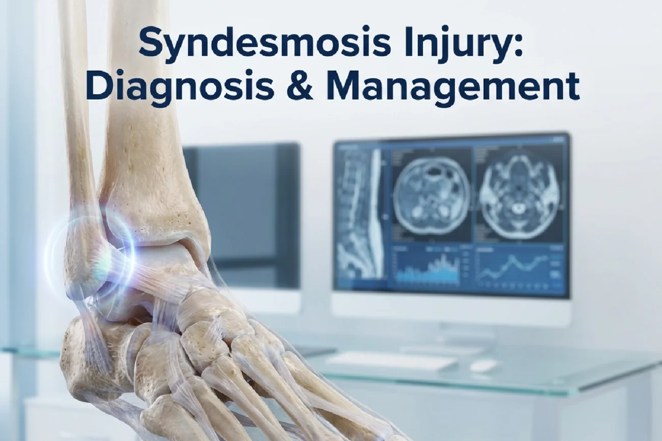
The distal tibiofibular syndesmosis is a complex, predominantly fibrous joint. It stabilises the ankle’s talar mortise, thereby facilitating controlled motion.
Syndesmosis injury occurs in up to 11% of all ankle sprains and approximately 10% of ankle fractures.
The primary cause is rotational trauma, specifically supination-external rotation and pronation-external rotation mechanisms. Therefore, doctors must accurately evaluate the degree of ligamentous injury and resulting instability. The joint comprises the distal tibia and fibula. Four key ligaments connect them, including the anterior and posterior tibiofibular ligaments. The transverse and interosseous ligaments complete this complex structure. Widening the ankle mortise by just 1 mm causes serious joint problems. In fact, this displacement decreases the tibiotalar joint contact area by 42%. Consequently, this can lead to early osteoarthritis. Furthermore, patients frequently present with complaints of persistent ankle instability months after an initial sprain.
Imaging Modalities for Syndesmotic Evaluation
Radiography is the first-line imaging modality. However, plain films and MRI have known limitations in reliably detecting instability under physiologic loads. Comparative weight-bearing radiographs, including anteroposterior, lateral, and mortise views, assist in initial diagnosis. Nevertheless, MRI remains the reference standard for comprehensively evaluating ligament integrity due to its high sensitivity. Emerging technologies, such as stress comparative CT and weight-bearing CT, offer substantial promise. These advanced modalities simulate physiologic loading conditions, thereby significantly improving the detection of subtle syndesmotic instability.
Management of Syndesmosis Injury and Reduction
Management strategies depend directly on the extent of the ligamentous injury and joint stability. Doctors generally treat patients with stable lesions conservatively, beginning with immobilisation for one to three weeks. Conversely, unstable syndesmosis injury typically requires surgical intervention. The primary goal of surgery is accurate syndesmosis reduction. This reduction is the strongest predictor of positive post-recovery outcomes. Surgical methods include syndesmotic screws, bioabsorbable material, and dynamic button-suture fixation techniques like TightRope. Also, physical examination is crucial for diagnosis before imaging. Specific clinical tests include the Squeeze (Hopkins) test and the external rotation stress test.
Frequently Asked Questions
Q1: What is the most critical factor for a positive outcome after surgical treatment?
Accurate reduction of the syndesmosis is the strongest predictor of a positive post-recovery outcome.
Q2: What are the four key ligaments that form the distal tibiofibular syndesmosis?
The complex is formed by the anterior tibiofibular ligament (ATIFL), the posterior tibiofibular ligament (PTIFL), the transverse ligament, and the interosseous ligament.
Q3: What imaging modality is the current reference standard for ligament integrity evaluation?
Magnetic Resonance Imaging (MRI) is the reference standard for evaluating the integrity of the syndesmotic ligaments, offering high sensitivity and specificity.
References
- Silva LNMD et al. Distal Tibiofibular Syndesmosis: Anatomy, Biomechanics, Imaging Approach, and Postoperative Evaluation. Radiographics. 2026 Feb undefined. doi: 10.1148/rg.250065. PMID: 41610035.
- Al-Tinawi et al. Management of Syndesmosis Injury: A Narrative Review. PMCID: PMC9745112. 2022 Dec 10. Available at: nih.gov. Accessed February 2, 2026.
- Munt et al. Anatomy of the distal tibiofibular syndesmosis in adults: a pictorial essay with a multimodality approach. Eur J Radiol. 2017 May. doi: 10.1016/j.ejrad.2017.02.007. Available at: nih.gov. Accessed February 2, 2026.
- Syndesmosis Injury Treatment – Melbourne. Orthotics Plus. Available at: orthoticsplus.com.au. Accessed February 2, 2026.