Can AI Replace Gadolinium Contrast in Routine Brain MRI?
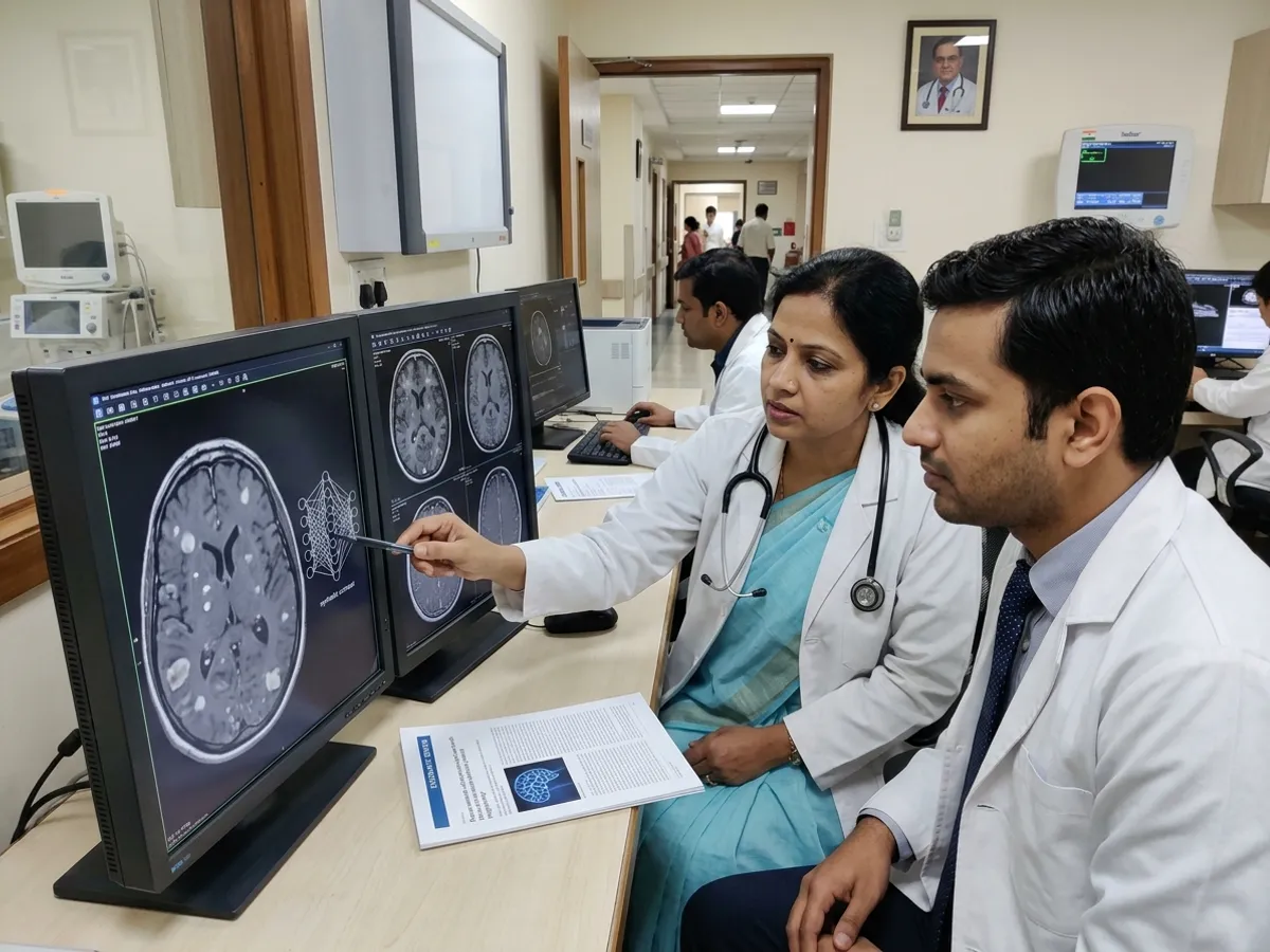
Gadolinium-based contrast agents play a pivotal role in diagnostic magnetic resonance imaging. However, concerns regarding tissue retention, renal tolerance, and acquisition costs have driven interest in artificial intelligence solutions. Consequently, researchers are evaluating synthetic postcontrast MRI to generate enhanced scans directly from unenhanced sequences. A new systematic review evaluates the technical accuracy and translational readiness of these deep learning models.
Current Landscape of Synthetic Postcontrast MRI
The comprehensive systematic review examined 41 adult studies investigating neural network architectures. Specifically, neuroimaging accounted for 59% of the literature, followed by breast imaging at 17% and body imaging at 15%. Generative adversarial networks and convolutional neural networks dominated the architectural designs.
Furthermore, most investigations evaluated image quality using whole-image metrics. Investigators reported the structural similarity index measure in 76% of papers and peak signal-to-noise ratio in 68% of papers. Therefore, these basic metrics showed strong technical feasibility on standard test sets.
Meta-Analysis Findings in Brain Tumor Imaging
The researchers performed a targeted random-effects meta-analysis across 15 brain tumor studies comprising 30 distinct models. Overall, the pooled structural similarity index measure reached 0.92, while the pooled peak signal-to-noise ratio averaged 30.6 decibels. In addition, these quantitative values suggest that algorithms reconstruct broad anatomical contours effectively.
However, the authors emphasized extreme statistical heterogeneity across the included literature. In particular, inconsistency in preprocessing workflows and metric computations limits direct comparisons. Moreover, pathology-specific evaluations revealed markedly lower fidelity within actual tumor boundaries than whole-brain metrics suggested.
Key Translational Barriers and Clinical Impact
Several practical barriers currently hinder clinical adoption. For instance, only 37% of reviewed studies conducted formal reader studies with radiologists. Similarly, 61% of models relied entirely on single-institution data, and only 29% shared their source code publicly. Consequently, algorithmic generalizability across different scanner vendors and pulse sequences remains largely unproven.
For clinicians, these findings provide important perspective. Although deep learning shows immense promise for contrast reduction, synthetic enhancement cannot yet replace real gadolinium in routine neuro-oncology. Thus, future research must prioritize external multicenter validation, standardized evaluation protocols, and task-specific clinical performance.
Frequently Asked Questions
Q1: What is synthetic postcontrast MRI?
Synthetic postcontrast MRI uses deep learning algorithms to predict contrast enhancement patterns from noncontrast sequences, eliminating or reducing the need for intravenous gadolinium agents.
Q2: Why do pathology-specific metrics matter more than whole-brain metrics?
Whole-brain metrics can artificially inflate performance because healthy tissue dominates the image. Conversely, lesion-specific metrics evaluate true diagnostic fidelity within abnormal tumor margins.
Q3: Is AI-generated contrast ready for routine patient care?
No, current models lack adequate external validation, multireader assessment, and standardized metrics. Therefore, synthetic imaging remains experimental until robust clinical trials establish safety.
References
- Dogra S et al. Deep Learning for Synthetic Postcontrast T1-Weighted MRI: A Systematic Review With Targeted Meta-Analysis of Brain Tumor Studies. AJR Am J Roentgenol. 2026 Aug 26. doi: 10.2214/AJR.26.34673. PMID: 42089523.
- Abbas S, Ali M, Ghalib L, et al. Virtual Gadolinium: Deep Learning-Based Virtual Contrast MRI Synthesis and Brain Tumor Segmentation. Spectr Eng Sci. 2026;2(1):45-58.
- Calabrese E, Rudie JD, Rauschecker AM, Villanueva-Meyer JE, Cha S. Feasibility of Simulated Postcontrast MRI of Glioblastomas and Lower-Grade Gliomas by Using Three-Dimensional Fully Convolutional Neural Networks. Radiol Artif Intell. 2021;3(5):e200276.