RaFiST: AI Radiomics Accurately Stratifies Lung Cancer

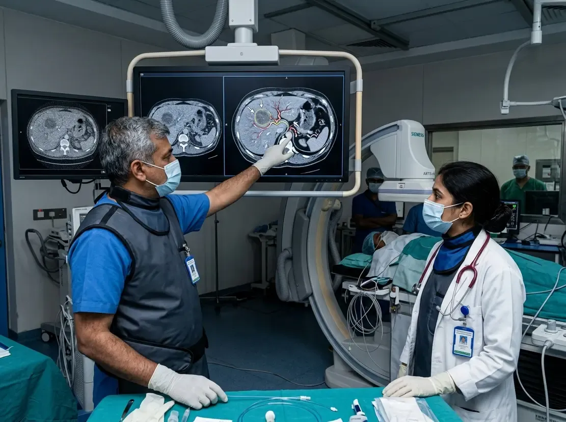
Assessing tumor fibrosis in NSCLC remains a vital step in understanding therapeutic resistance and disease aggressiveness. Intratumoral collagen deposition often impedes effective drug delivery and alters the immune microenvironment. However, conventional histopathological evaluation requires invasive tissue biopsy, which is not always feasible. To address this challenge, researchers recently introduced RaFiST, an innovative artificial intelligence framework. This novel tool non-invasively evaluates tumor collagen levels using routine contrast-enhanced computed tomography (CT) scans.
How RaFiST Measures Tumor Fibrosis in NSCLC
The multicenter retrospective investigation evaluated 532 patients with surgically resected lung cancer across two clinical centers. Pathologists first quantified tumor collagen fraction using digital image analysis on stained tissue sections. Next, investigators extracted high-dimensional radiomic features from preoperative CT images. The team then developed the RaFiST signature to capture subtle imaging textures corresponding to dense stromal matrices.
Importantly, the model demonstrated strong diagnostic capability. RaFiST achieved an area under the curve (AUC) of 0.879 in the training cohort. Furthermore, it maintained robust accuracy with an AUC of 0.813 in an independent external test cohort. Using an optimal histopathological cutoff of 11.12%, the model effectively divided patients into high- and low-fibrosis risk categories.
Prognostic Significance and Clinical Implications
Multivariable analysis confirmed that the intratumoral fibrosis score serves as an independent predictor of disease-free survival and overall survival. Specifically, patients classified into the high-fibrosis group experienced significantly worse clinical outcomes. Therefore, integrating radiomics-based stroma assessment into routine oncology workflows can enhance risk stratification. Moreover, the combined model outperformed standard clinical staging systems alone. Transcriptomic analyses also revealed distinct molecular pathways driving this aggressive fibrotic phenotype, highlighting the necessity for specialized education like a Certification Course In Lung Cancer to better manage advanced malignancies.
Frequently Asked Questions
Q1: What is the clinical significance of tumor fibrosis in NSCLC?
Tumor fibrosis promotes therapeutic resistance and shields malignant cells from immune surveillance. Consequently, high collagen deposition correlates with shorter disease-free and overall survival in lung cancer patients, areas extensively covered in Certification Course In Clinical Oncology programs.
Q2: How does the RaFiST model quantify fibrosis non-invasively?
RaFiST extracts quantitative texture and morphological features from preoperative contrast-enhanced CT scans. Thus, it accurately estimates histological collagen volume without requiring additional invasive tissue sampling.
References
- Zong Y et al. RaFiST: a radiomics model for non-invasive stratification of tumor fibrosis and prognostic prediction in non-small cell lung cancer. Eur Radiol. 2026 Aug 20. doi: 10.1007/s00330-026-12764-5. PMID: 42622885.
- Chen S et al. Radiomics reveals the biological basis for non-small cell lung cancer prognostic stratification by reflecting tumor immune microenvironment heterogeneity. Front Immunol. 2025;16:1708692.
- Khorrami R et al. Radiomics-Based Prediction of Treatment Response in Non-Small Cell Lung Cancer Using Pre-Treatment CT Imaging Features. MDPI. 2026.