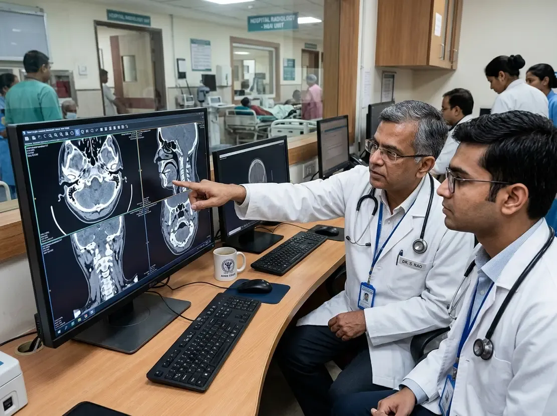
Predicting ESCC Outcomes with Nodal Imaging Features

Predicting ESCC Outcomes with Nodal Imaging Features
Researchers recently explored Esophageal Cancer Risk Stratification techniques to improve patient outcomes after surgery. Specifically, they looked at how lymph node changes during treatment can predict future recurrence. This study focuses on patients with locally advanced esophageal squamous cell carcinoma receiving neoadjuvant immunochemotherapy. Consequently, these findings offer a new way to personalize follow-up care for Indian patients facing high ESCC prevalence.
Esophageal Cancer Risk Stratification via Imaging
The study analyzed temporal imaging features of the largest lymph node after treatment. Researchers found that certain imaging characteristics serve as independent predictors of positive lymph nodes. Furthermore, these findings allow doctors to identify high-risk subgroups even among those who appear to have negative nodes pathologically. Therefore, clinicians can make better decisions regarding adjuvant therapy. These insights help bridge the gap between imaging data and clinical pathology.
Improved Prognostic Models for Clinical Use
The team constructed a predictive model using significant imaging variables. Their results showed high accuracy in both training and validation cohorts. Additionally, the model effectively stratified patients into high-risk and low-risk groups. Patients in the high-risk group experienced significantly shorter disease-free survival. Thus, this imaging-based approach offers a reliable way to monitor treatment response. Moreover, it provides a non-invasive method to refine prognosis before surgical intervention.
Frequently Asked Questions
Q1: How does temporal imaging help in ESCC management?
Temporal imaging tracks changes in lymph nodes during neoadjuvant therapy. These changes provide vital clues about the tumor’s response to immunotherapy. Consequently, doctors can predict which patients might face a higher risk of recurrence after surgery.
Q2: Why is identifying high-risk ypN0 patients important?
Some patients have pathologically negative nodes but still face a high risk of cancer returning. Identifying these individuals allows for more aggressive adjuvant therapy. Therefore, this stratified approach improves long-term survival chances for esophageal cancer patients.
References
- Fan T et al. Temporal lymph node imaging features for prognostic risk stratification in neoadjuvant immunotherapy for esophageal cancer: a multicenter study. Eur Radiol. 2026 Apr 27. doi: 10.1007/s00330-026-12566-9. PMID: 42045511.
- Kelly RJ et al. Adjuvant Nivolumab in Resected Esophageal or Gastroesophageal Junction Cancer. N Engl J Med. 2021;384(13):1191-1203.
- Sung H et al. Global Cancer Statistics 2020: GLOBOCAN Estimates of Incidence and Mortality Worldwide for 36 Cancers in 185 Countries. CA Cancer J Clin. 2021;71(3):209-249.