Non-Contrast MRI Shows Promise for Endolymphatic Hydrops
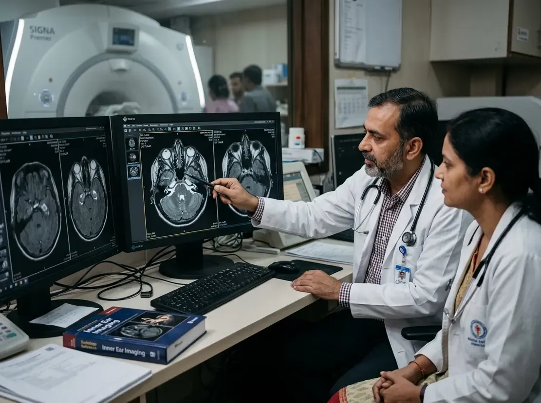
Clinicians often face challenges when diagnosing Ménière's disease and inner ear pathology. Currently, delayed contrast-enhanced scans serve as the clinical standard. However, an innovative unenhanced endolymphatic hydrops MRI technique may transform this diagnostic pathway. This novel method utilizes three-dimensional fluid-attenuated inversion recovery sequences acquired at two different inversion times. Furthermore, digital subtraction of these sequences highlights anatomical fluid compartments without administering gadolinium-based agents.
The Mechanism of Endolymphatic Hydrops MRI Subtraction
The inner ear contains two distinct fluid spaces, namely the perilymph and the endolymph. Traditionally, clinicians rely on delayed intravenous gadolinium to differentiate these microstructures. The contrast agent distributes exclusively into the perilymph after four hours. In contrast, the unenhanced technique leverages the intrinsic relaxation differences between the two fluids.
Researchers demonstrated that endolymphatic and perilymphatic signals exhibit distinct signal intensity trajectories across different inversion times. Consequently, imaging at two targeted inversion times enables signal separation. Clinicians then subtract the shorter inversion time image from the longer inversion time image. Therefore, this subtraction selectively suppresses perilymph or endolymph signal. The resulting image visualizes vestibular and cochlear hydrops without any chemical contrast exposure.
Clinical Validation and Diagnostic Performance
A recent clinical evaluation assessed this subtraction technique across 202 patients. The investigators compared the unenhanced subtraction images with standard enhanced 3D-FLAIR and 3D-real inversion-recovery scans. Notably, the unenhanced images yielded diagnostic features remarkably similar to gadolinium-enhanced 3D-real IR.
Although the unenhanced subtraction protocol produced lower subjective image quality scores, diagnostic concordance remained notably high. Specifically, observers achieved substantial agreement when grading cochlear and vestibular endolymphatic hydrops. Moreover, the technique reliably identified clinically significant dilation in patients presenting with vertigo. As a result, this non-invasive approach provides a dependable screening alternative for symptomatic patients.
Significance for Clinical Practice in India
This development holds tremendous practical significance for neurotology and radiology practices in India. Standard delayed contrast imaging requires patients to wait four hours between intravenous injection and scanning. Consequently, this lengthy waiting period strains outpatient radiology workflows and burdens patients traveling from rural areas.
In addition, gadolinium administration adds substantial financial cost for self-paying individuals. Contrast agents also present safety concerns in patients with severe renal dysfunction or gadolinium hypersensitivity. Therefore, eliminating contrast media avoids adverse events and reduces direct healthcare expenditures. Indian centers can implement these subtraction sequences on modern 3 Tesla MRI scanners without specialized hardware. Thus, this unenhanced method broadens patient access to definitive vestibular diagnostics.
Frequently Asked Questions
Q1: Why is unenhanced endolymphatic hydrops MRI important?
Unenhanced imaging eliminates the need for intravenous gadolinium, which reduces adverse reaction risks and cuts procedural costs. Furthermore, it avoids the traditional four-hour post-injection waiting delay, streamlining clinic workflow.
Q2: How does 3D-FLAIR subtraction separate inner ear fluids?
Perilymph and endolymph exhibit different signal recovery rates across varying inversion times. Therefore, subtracting an image acquired at a shorter inversion time from one with a longer inversion time isolates the fluid compartments clearly.
Q3: Can this method fully replace contrast-enhanced MRI in Ménière's disease?
Currently, the method shows strong diagnostic concordance but slightly lower image quality than delayed contrast scans. Consequently, clinicians may utilize it as a practical screening alternative, especially when contrast media are contraindicated.
References
- Zhao M et al. Enabling unenhanced endolymphatic hydrops MRI: a subtraction method using 3D-FLAIR sequences with different inversion time. Eur Radiol. 2026 Oct 08. doi: 10.1007/s00330-026-12930-9. PMID: 42848041.
- Baráth K, Schuknecht B, Naldi AM, Schrepfer T, Bockisch CJ, Hegemann SC. Detection and grading of endolymphatic hydrops in Menière disease using MR imaging. AJNR Am J Neuroradiol. 2014;35(7):1387-1392.
- Bernasconi N, Bernaerts A, Casselman J, et al. Detailed insight into magnetic resonance assessment of Ménière's disease – description of methodology and imaging findings in a case series. Pol J Radiol. 2022;87:354-362.