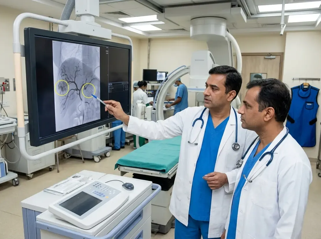
AI Transforms Pediatric Kidney Imaging: UroVision Study
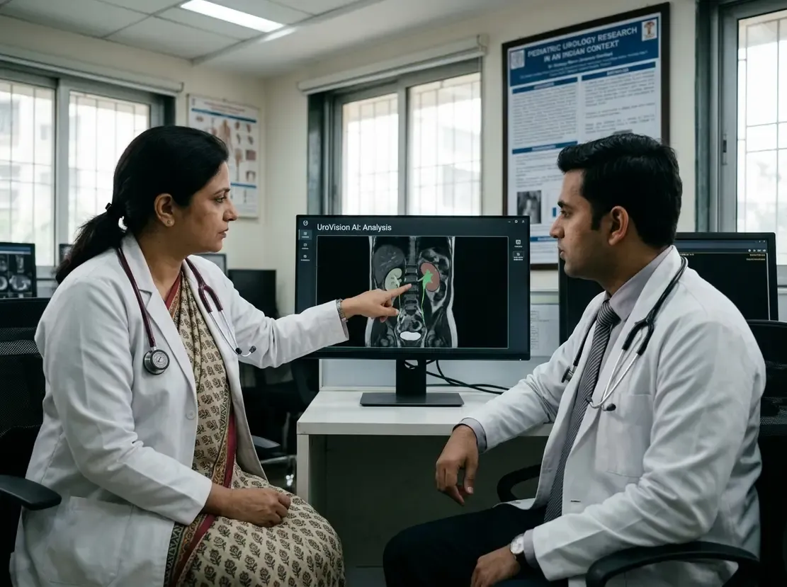
Congenital anomalies of the kidney and urinary tract represent the leading cause of pediatric chronic kidney disease worldwide. Therefore, timely and accurate structural and functional evaluation remains essential to safeguard nephron mass. Clinicians increasingly favor functional MR urography because it delivers comprehensive dynamic assessment without ionizing radiation. However, traditional manual and semi-automated post-processing workflows demand significant time and operator expertise. In response, investigators developed UroVision, a novel artificial intelligence tool designed for automated segmentation and dynamic analysis. This innovation addresses workflow bottlenecks while promoting standardized diagnostic decisions across pediatric urology and radiology units.
Advancing Functional MR Urography with AI Automation
The validation study compared UroVision against the established ImageJ MRU platform across fifty pediatric patients with confirmed congenital anomalies. Consequently, the researchers analyzed one hundred kidneys to determine diagnostic agreement and operational reliability. Two pediatric radiologists independently evaluated split renal function, parenchymal volumes, and urinary drainage curves. To maintain consistency with ImageJ MRU, the primary functional evaluation examined the two-dimensional slice containing the greatest renal parenchyma. Furthermore, the expert team reviewed full three-dimensional UroVision data to resolve ambiguous or borderline clinical findings. This methodological comparison established clear operational benchmarks for clinical translation.
Robust Performance and High Reproducibility
The study yielded impressive quantitative results across all key functional and anatomical metrics. Specifically, renal volume measurements showed near-perfect correlation between the two platforms at r = 0.99. In addition, functional indicators demonstrated moderate correlation, including glomerular slope and Patlak coefficient values. Bland-Altman analysis revealed minimal systematic bias between the platforms. Moreover, reader reproducibility proved exceptional, showing intraclass correlation coefficients above 0.87 for both inter-reader and intra-reader evaluations. Qualitative urinary drainage curve classification also demonstrated strong concordance, yielding a weighted Cohen kappa of 0.86. Importantly, full three-dimensional analysis successfully reclassified borderline drainage curves in sixteen out of twenty-three kidneys.
Clinical Implications for Pediatric Practice
These findings carry significant relevance for pediatric nephrologists, urologists, and radiologists caring for children with congenital uropathies. Historically, complex post-processing restricted comprehensive dynamic magnetic resonance studies to specialized tertiary centers. Because UroVision automates three-dimensional parenchymal segmentation, it substantially minimizes operator dependence and post-processing times. Thus, busy clinical units can deploy standardized functional urography more readily into routine clinical pathways. In practice, automated split renal function and drainage categorization assist clinicians in detecting obstruction and selecting timely surgical intervention. Ultimately, adopting automated analytical platforms helps clinicians preserve kidney function while avoiding repetitive nuclear scintigraphy examinations.
Frequently Asked Questions
Q1: What is the main clinical advantage of UroVision over existing software?
UroVision provides fully automated three-dimensional segmentation and functional analysis. Consequently, it minimizes user-dependent variations and shortens post-processing time compared to manual platforms.
Q2: How well does UroVision agree with established ImageJ MRU results?
The platform shows near-perfect agreement for renal volume calculation and high concordance for urinary drainage curves. Furthermore, it demonstrates excellent inter-reader and intra-reader reproducibility.
Q3: How does three-dimensional analysis improve clinical decision-making in CAKUT?
Full three-dimensional analysis resolves equivocal two-dimensional findings. For example, it successfully clarified borderline drainage curves in the majority of indeterminate kidneys.
References
- Gnocchi G et al. UroVision: a new artificial intelligence software for functional magnetic resonance urography evaluation in children with congenital anomalies of the kidney and urinary tract. Eur Radiol. 2026 Oct 08. doi: 10.1007/s00330-026-12950-5. PMID: 42848042.
- Damasio MB et al. Comparative Study Between Functional MR Urography and Renal Scintigraphy to Evaluate Drainage Curves and Split Renal Function in Children With Congenital Anomalies of Kidney and Urinary Tract (CAKUT). Front Pediatr. 2020;7:527. doi: 10.3389/fped.2019.00527.
- Press B, Germany R, Coronado M, Kirsch A. Pediatric Magnetic Resonance Urography: Emerging Technologies and the Future of Quantitative Renal Imaging. Med Res Arch. 2025;13(10). doi: 10.18103/mra.v13i10.6990.